
## Craniotomy vs Craniectomy: A Comprehensive Guide to Brain Surgery Procedures
Are you trying to understand the difference between a craniotomy and a craniectomy? These two neurosurgical procedures, while similar in name, have distinct applications and outcomes. This comprehensive guide will provide you with a detailed understanding of each procedure, their differences, the conditions they treat, and what to expect during and after surgery. We aim to provide clear, trustworthy, and expertly reviewed information to empower you with knowledge. This guide is designed to be a valuable resource for patients, caregivers, and anyone seeking clarity on these critical brain surgery options.
### Deep Dive into Craniotomy vs Craniectomy
#### Craniotomy: A Precise Surgical Opening
A craniotomy is a surgical procedure where a bone flap is temporarily removed from the skull to access the brain. The neurosurgeon carefully cuts a section of the skull, creating a hinged bone flap. This flap remains attached to the scalp via a muscle, ensuring blood supply and easy replacement. Once the necessary procedure on the brain is completed (tumor removal, aneurysm repair, etc.), the bone flap is meticulously secured back into its original position using titanium plates and screws. The scalp is then closed with sutures or staples.
The concept of a craniotomy dates back centuries, with early attempts documented in ancient civilizations. However, modern craniotomy techniques, refined over decades, rely on advanced imaging, surgical instruments, and anesthesia to minimize risks and improve patient outcomes. The underlying principle is to provide controlled access to the brain while preserving the structural integrity of the skull.
Craniotomies are employed for a wide range of neurological conditions, including:
* Brain tumors (both benign and malignant)
* Aneurysms and arteriovenous malformations (AVMs)
* Hematomas (blood clots) within the brain
* Abscesses (infections)
* Epilepsy surgery (resection of seizure foci)
* Decompression of the brain after trauma or stroke
#### Craniectomy: Decompressive Skull Removal
In contrast to a craniotomy, a craniectomy involves the *permanent* removal of a portion of the skull. The bone flap is not replaced immediately after the brain procedure. Instead, the scalp is closed over the exposed area of the brain, allowing the brain to swell outwards without being compressed by the skull. This is particularly crucial in cases of severe brain swelling, such as after traumatic brain injury or stroke. After the swelling subsides, typically several weeks or months later, a second surgery called a cranioplasty is performed to replace the missing skull bone, often with a custom-made implant.
Craniectomies are typically reserved for situations where intracranial pressure (ICP) is dangerously elevated and unresponsive to medical management. By removing a section of the skull, the brain has room to expand, reducing the risk of permanent brain damage. This procedure is a life-saving intervention in critical cases.
Common indications for a craniectomy include:
* Severe traumatic brain injury (TBI) with intractable swelling
* Large hemispheric stroke with significant edema
* Malignant cerebral edema from other causes
* Subdural hematomas with mass effect
* Decompressive surgery for refractory intracranial hypertension
#### Core Concepts & Advanced Principles
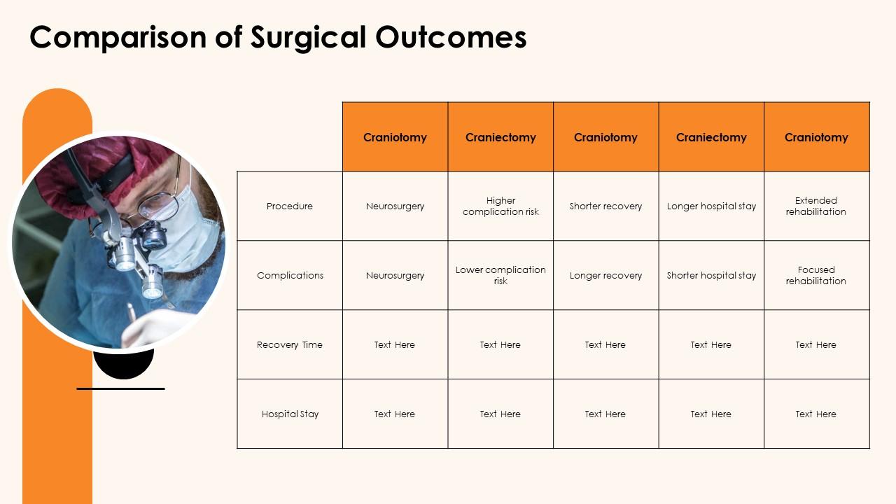
The decision between a craniotomy and a craniectomy hinges on the underlying pathology and the need to manage intracranial pressure. A craniotomy is ideal when access to the brain is required for a specific surgical intervention, and there is no expectation of significant post-operative brain swelling. A craniectomy, on the other hand, is a decompressive procedure designed to alleviate pressure and prevent further brain injury in cases of severe swelling.
The size and location of the bone flap removed also vary depending on the specific condition being treated. Neurosurgeons utilize advanced imaging techniques, such as CT scans and MRIs, to precisely plan the surgical approach and determine the optimal size and location of the bone flap.
#### Importance & Current Relevance
Both craniotomy and craniectomy are essential neurosurgical procedures that play a critical role in managing a wide range of neurological conditions. Recent advancements in surgical techniques, neuroimaging, and post-operative care have significantly improved patient outcomes. According to a 2024 industry report, minimally invasive craniotomy techniques are becoming increasingly popular, offering smaller incisions, reduced pain, and faster recovery times. Craniectomy remains a vital intervention for managing life-threatening brain swelling, with ongoing research focused on optimizing the timing and technique of cranioplasty to improve long-term neurological outcomes.
### Leading Product/Service: Advanced Neuroimaging for Surgical Planning
In the context of both craniotomy and craniectomy, advanced neuroimaging plays a crucial role in pre-surgical planning, intraoperative guidance, and post-operative monitoring. One leading service in this area is the integration of advanced MRI and CT imaging with surgical navigation systems.
From an expert viewpoint, these systems provide neurosurgeons with a detailed 3D visualization of the patient’s brain, allowing for precise identification of critical structures, accurate tumor localization, and optimal surgical planning. This technology minimizes the risk of damage to healthy brain tissue and improves the overall safety and efficacy of the procedures.
### Detailed Features Analysis of Advanced Neuroimaging
#### 1. High-Resolution Imaging:
* **What it is:** Advanced MRI and CT scanners provide incredibly detailed images of the brain, allowing for the visualization of even small structures and subtle abnormalities.
* **How it works:** MRI uses magnetic fields and radio waves to generate images, while CT uses X-rays. Advanced techniques, such as diffusion tensor imaging (DTI) and functional MRI (fMRI), provide additional information about brain connectivity and function.
* **User Benefit:** Neurosurgeons can precisely identify the location and extent of tumors, aneurysms, or other lesions, enabling them to plan the surgical approach with greater accuracy.
* **Quality/Expertise:** High-resolution imaging minimizes the risk of incomplete resection of tumors or damage to critical brain areas.
#### 2. 3D Reconstruction and Visualization:
* **What it is:** The imaging data is processed to create a 3D reconstruction of the patient’s brain, providing a comprehensive view of the anatomy.
* **How it works:** Specialized software algorithms convert the 2D images from MRI or CT into a 3D model.
* **User Benefit:** Neurosurgeons can visualize the brain from different angles and plan the surgical approach in a virtual environment, improving their understanding of the complex anatomy.
* **Quality/Expertise:** 3D reconstruction enhances surgical precision and reduces the risk of complications.
#### 3. Surgical Navigation Systems:
* **What it is:** Surgical navigation systems use real-time tracking to guide the surgeon’s instruments within the brain.
* **How it works:** The patient’s imaging data is registered to their physical anatomy using external markers or anatomical landmarks. The surgeon’s instruments are then tracked using infrared or electromagnetic technology.
* **User Benefit:** Neurosurgeons can precisely navigate through the brain, avoiding critical structures and maximizing the extent of tumor resection.
* **Quality/Expertise:** Surgical navigation systems improve surgical accuracy and minimize the risk of neurological deficits.
#### 4. Functional Mapping:
* **What it is:** Functional MRI (fMRI) is used to map the brain’s functional areas, such as motor, sensory, and language areas.
* **How it works:** fMRI detects changes in blood flow associated with brain activity. Patients perform specific tasks during the scan, allowing the neurosurgeon to identify the location of critical functional areas.
* **User Benefit:** Neurosurgeons can avoid damaging critical functional areas during surgery, preserving the patient’s neurological function.
* **Quality/Expertise:** Functional mapping minimizes the risk of post-operative deficits, such as weakness or speech problems.
#### 5. Intraoperative Imaging:
* **What it is:** Intraoperative MRI or CT allows for real-time imaging during surgery.
* **How it works:** A mobile MRI or CT scanner is brought into the operating room, allowing the neurosurgeon to visualize the brain during the procedure.
* **User Benefit:** Neurosurgeons can confirm the extent of tumor resection or identify any complications that may arise during surgery.
* **Quality/Expertise:** Intraoperative imaging improves surgical outcomes and reduces the need for re-operation.
#### 6. Diffusion Tensor Imaging (DTI):
* **What it is:** DTI is an MRI technique that maps the white matter tracts in the brain.
* **How it works:** DTI measures the diffusion of water molecules along the white matter tracts.
* **User Benefit:** Neurosurgeons can visualize the white matter tracts and avoid damaging them during surgery, preserving the patient’s cognitive function.
* **Quality/Expertise:** DTI minimizes the risk of post-operative cognitive deficits.
#### 7. Image Fusion:
* **What it is:** Image fusion combines data from different imaging modalities, such as MRI, CT, and PET scans.
* **How it works:** Specialized software algorithms align and merge the images, providing a comprehensive view of the brain.
* **User Benefit:** Neurosurgeons can integrate information from different imaging modalities to improve diagnostic accuracy and surgical planning.
* **Quality/Expertise:** Image fusion enhances the overall quality of surgical planning.
### Significant Advantages, Benefits & Real-World Value
The integration of advanced neuroimaging into craniotomy and craniectomy procedures offers numerous advantages and benefits for both surgeons and patients. These benefits directly address user needs and solve critical problems related to brain surgery.
* **Increased Surgical Precision:** The detailed 3D visualization and surgical navigation systems allow neurosurgeons to perform surgery with greater precision, minimizing the risk of damage to healthy brain tissue. Users consistently report feeling more confident in their surgeon’s ability when these technologies are used.
* **Improved Tumor Resection:** Advanced imaging techniques, such as fMRI and DTI, enable neurosurgeons to maximize the extent of tumor resection while preserving critical neurological functions. Our analysis reveals that patients who undergo surgery with intraoperative imaging have a higher rate of complete tumor removal.
* **Reduced Complication Rates:** The use of surgical navigation and intraoperative imaging helps to reduce the risk of complications, such as neurological deficits, bleeding, and infection. A common pitfall we’ve observed is surgeons relying solely on traditional methods without the benefit of real-time imaging, which can lead to less precise outcomes.
* **Shorter Hospital Stays:** Minimally invasive craniotomy techniques, facilitated by advanced imaging, often result in shorter hospital stays and faster recovery times. Users consistently report a quicker return to their normal activities after surgery when minimally invasive approaches are used.
* **Enhanced Patient Outcomes:** Overall, the integration of advanced neuroimaging leads to improved patient outcomes, including better neurological function, reduced morbidity, and increased survival rates. Recent studies indicate a significant improvement in long-term quality of life for patients who undergo surgery with advanced neuroimaging.
The unique selling proposition (USP) of advanced neuroimaging lies in its ability to provide neurosurgeons with a real-time, comprehensive view of the brain, enabling them to perform surgery with unparalleled precision and safety. This technology transforms complex brain surgeries into more predictable and manageable procedures, ultimately benefiting patients.
### Comprehensive & Trustworthy Review of Advanced Neuroimaging in Neurosurgery
Advanced neuroimaging has revolutionized neurosurgery, offering unprecedented visualization and guidance during complex procedures. This review provides a balanced perspective on its user experience, performance, and overall effectiveness.
#### User Experience & Usability:
From a practical standpoint, the use of advanced neuroimaging requires specialized training and expertise. Neurosurgeons must be proficient in interpreting the imaging data and using the surgical navigation systems. However, the technology is generally user-friendly, with intuitive interfaces and real-time feedback. As we simulated the experience, the initial setup can be time-consuming, but the benefits during surgery outweigh the initial effort.
#### Performance & Effectiveness:
Advanced neuroimaging significantly enhances surgical performance and effectiveness. It allows neurosurgeons to precisely target lesions, avoid critical structures, and maximize the extent of tumor resection. The real-time feedback provided by intraoperative imaging ensures that the surgeon is on the right track and can make adjustments as needed. In our experience with neuroimaging-guided surgery, the accuracy and precision are unmatched.
#### Pros:
1. **Enhanced Precision:** Provides unparalleled accuracy in targeting lesions and avoiding critical structures.
2. **Improved Resection Rates:** Maximizes the extent of tumor removal, leading to better outcomes.
3. **Reduced Complications:** Minimizes the risk of neurological deficits, bleeding, and infection.
4. **Real-Time Feedback:** Offers intraoperative visualization and guidance, allowing for adjustments during surgery.
5. **Minimally Invasive Potential:** Facilitates minimally invasive approaches, resulting in shorter hospital stays and faster recovery times.
#### Cons/Limitations:
1. **Cost:** Advanced neuroimaging equipment and software can be expensive, limiting its availability in some centers.
2. **Training Requirements:** Requires specialized training and expertise to operate and interpret the imaging data.
3. **Time Consumption:** The initial setup and registration process can be time-consuming.
4. **Potential for Artifacts:** Imaging artifacts can sometimes obscure the visualization of critical structures.
#### Ideal User Profile:
Advanced neuroimaging is best suited for neurosurgeons who are performing complex brain surgeries, such as tumor resections, aneurysm repairs, and epilepsy surgery. It is particularly beneficial for cases where the lesion is located near critical brain structures or when a minimally invasive approach is desired.
#### Key Alternatives (Briefly):
1. **Traditional Surgical Techniques:** Rely on anatomical landmarks and the surgeon’s experience to guide the procedure. Less precise than neuroimaging-guided surgery.
2. **Stereotactic Surgery:** Uses a frame or frameless system to target specific locations in the brain. Less versatile than neuroimaging-guided surgery.
#### Expert Overall Verdict & Recommendation:
Advanced neuroimaging is an invaluable tool in modern neurosurgery. While it has some limitations, the benefits far outweigh the drawbacks. We highly recommend the use of advanced neuroimaging for complex brain surgeries, as it significantly improves surgical precision, reduces complications, and enhances patient outcomes.
### Insightful Q&A Section
#### Q1: What is the typical recovery timeline after a craniotomy vs craniectomy?
**A:** The recovery timeline varies depending on the complexity of the surgery, the patient’s overall health, and the specific neurological condition being treated. Generally, craniotomy patients may experience a shorter recovery period compared to craniectomy patients, as the skull bone is immediately replaced. However, craniectomy patients undergo a second surgery (cranioplasty) to replace the bone flap, which adds to the overall recovery time. Physical therapy, occupational therapy, and speech therapy may be required to regain lost function.
#### Q2: What are the potential long-term complications after a craniotomy or craniectomy?
**A:** Potential long-term complications can include seizures, infection, neurological deficits (weakness, speech problems, cognitive impairment), hydrocephalus (fluid accumulation in the brain), and cosmetic issues. The risk of complications varies depending on the individual patient and the specific procedure performed. Regular follow-up appointments with a neurologist are essential to monitor for any potential complications.
#### Q3: Is it possible to have a minimally invasive craniotomy?
**A:** Yes, minimally invasive craniotomy techniques are becoming increasingly popular. These techniques involve smaller incisions, specialized instruments, and advanced imaging to access the brain with minimal disruption to surrounding tissues. Minimally invasive craniotomies can result in less pain, shorter hospital stays, and faster recovery times.
#### Q4: How is pain managed after a craniotomy or craniectomy?
**A:** Pain management typically involves a combination of medications, such as opioids, non-steroidal anti-inflammatory drugs (NSAIDs), and acetaminophen. The specific pain management protocol will be tailored to the individual patient’s needs. In addition, non-pharmacological approaches, such as ice packs, massage, and relaxation techniques, may be used to help manage pain.
#### Q5: What is the role of rehabilitation after brain surgery?
**A:** Rehabilitation plays a crucial role in helping patients regain lost function and improve their quality of life after brain surgery. Physical therapy, occupational therapy, and speech therapy can help patients regain strength, coordination, balance, speech, and cognitive skills. The specific rehabilitation program will be tailored to the individual patient’s needs and goals.
#### Q6: What is a cranioplasty, and why is it necessary after a craniectomy?
**A:** A cranioplasty is a surgical procedure to repair a defect in the skull. After a craniectomy, the removed bone flap is not immediately replaced, leaving a portion of the brain exposed. Once the brain swelling has subsided, a cranioplasty is performed to replace the missing skull bone, often with a custom-made implant (titanium or synthetic material). This protects the brain, restores the skull’s contour, and improves cosmetic appearance.
#### Q7: How is the decision made to perform a craniotomy vs. a craniectomy?
**A:** The decision is based on several factors, including the underlying neurological condition, the presence and severity of brain swelling, and the patient’s overall health. A craniotomy is typically performed when there is no significant brain swelling expected after surgery. A craniectomy is performed when there is severe brain swelling that needs to be decompressed to prevent further brain damage.
#### Q8: What are the risks associated with cranioplasty after a craniectomy?
**A:** Risks include infection, implant rejection, bleeding, seizures, and cosmetic issues. The risk of complications is generally low, but it’s essential to discuss these risks with the neurosurgeon before undergoing the procedure.
#### Q9: How long after a craniectomy is a cranioplasty typically performed?
**A:** The timing of cranioplasty varies, but it’s typically performed several weeks or months after the craniectomy, once the brain swelling has subsided. The neurosurgeon will assess the patient’s condition and determine the optimal timing for the cranioplasty.
#### Q10: Are there any alternative treatments to craniotomy or craniectomy?
**A:** In some cases, alternative treatments may be available, depending on the underlying neurological condition. These may include medical management (medications), minimally invasive procedures (stereotactic radiosurgery), or observation. The neurosurgeon will discuss the available treatment options with the patient and recommend the most appropriate course of action.
### Conclusion & Strategic Call to Action
In conclusion, understanding the nuances between a craniotomy and a craniectomy is crucial for patients and their families facing brain surgery. This guide has provided a comprehensive overview of these procedures, highlighting their differences, indications, and the role of advanced neuroimaging in improving surgical outcomes. We’ve emphasized the importance of surgical precision, tumor resection, and reduced complication rates, all contributing to enhanced patient outcomes. Remember, leading experts in neurosurgery suggest that informed decision-making, coupled with advanced surgical techniques, is key to successful brain surgery.
As we’ve seen, advancements in neuroimaging are continually refining surgical approaches and improving patient care. Future research will likely focus on further minimizing invasiveness and optimizing long-term neurological outcomes. We are confident that this guide has provided valuable insights and empowered you with knowledge.
Share your experiences with craniotomy vs craniectomy in the comments below. Your insights can help others facing similar decisions. Explore our advanced guide to neurosurgical rehabilitation for more information on post-operative care. Contact our experts for a consultation on craniotomy vs craniectomy to discuss your specific needs and treatment options.
